
Genetic analysis of the brain development and function using zebrafish as a model system
- Analysis of genes involved in development of identifiable neurons and their functions in animal behaviors
- Live imaging of the neural circuit formation
- Visualizing brain morphogenesis
- Analysis of mutants
- Application of techniques invented to mammals for comparison
- Optogenetic analysis of neurons and muscles using ChR2
- High-resolution micro-CT and cinematography of neural crest-derived organ, pharyngeal teeth
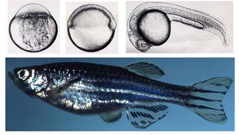
Zebrafish are small tropical fish whose body length is about 3 cm. Since their embryos are transparent and develop very rapidly, it is possible to observe brain and other organ formation in a living animal at the single cell resolution. It is also possible to screen mutants using ENU or gamma-ray methods. We will elucidate ‘the common basic mechanisms’ in vertebrates for brain differentiation, morphogenesis and evolution by identifying and visualizing important genes, neurons and circuits.
Zebrafish brain and Mauthner neurons
Zebrafish brain is much simpler than any other vertebrates, but its basic structure is very similar. For example, presence of the forebrain, midbrain and hindbrain, as well as the repeated segments in the hindbrain is a common feature shared with birds or mammals. Neurons involved in each particular function develop in a specific segment. For this purpose, a variety of transcriptional factors including homeo-domain proteins, and signaling molecules as well as cell surface molecules are involved.
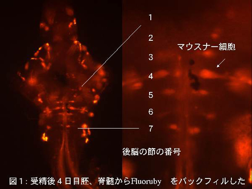
From the center of the 4th hindbrain segment, Mauthner cells (M-cells) , a pair of the biggest neurons in the brain, named after its discoverer, are developed. When the fish encounters a danger situation, M- cells collect a variety of sensory inputs to judge, and send signals to motoneurons that can contract body muscles in about 10 milliseconds to escape.
Fig: Dorsal view of the zebrafish brain (5 day larvae) backfilled with Fluoruby
Research using Mauthner cells as a model system
We will study gene function in the Mauthner development and function, by identifying novel genes. By focusing on an identifiable neuron as a model system, we hope to identify new mechanisms that may be applicable to other brain regions also.
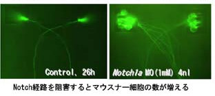
Molecular mechanism to specify the number of Mauthner neurons that are only present as a pair in the brain
Fig: There are only two M-cells in a brain. Thus it is possible to conduct research in details at high resolution. We have been running a frontier of their research using genetic, neural circuit formation as well as electro-physiological methods. When Delta-Notch pathway is blocked, we could obtain animals with have numerous M-cells, and they could be used for various research.
Visualization of neurons using fluorescent proteins
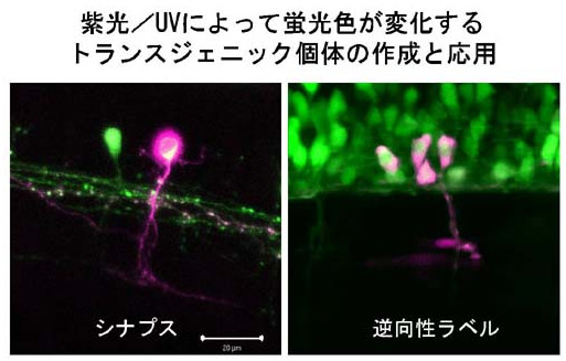
Creation of transgenic animals whose fluorescent color could be changed upon irradiation with UV
We succeeded in visualizing individual neuronal pathways in a brain of living animal using a fluorescent protein called Kaede. Kaede has a structure similar to GFP, but its color could be irreversibly changed from green to red upon irradiation with UV or 405nm laser.
Fig: Synapses; Retrograde labeling
Tracing morphogenetic movement
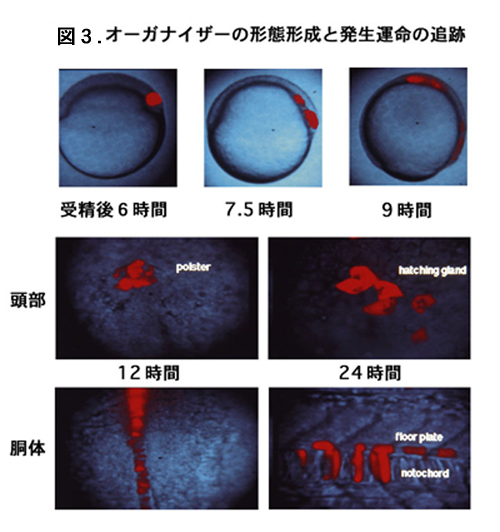
We also looking for morphogenetic movement and tissue interactions immediately before the neural differentiation.
Fig.3: Morphogenesis and the developmental fate of the organizer.